Microbiology laboratory manuals, often available as PDF resources, detail essential techniques and principles. These guides cover staining, culturing, and biochemical tests for bacterial identification, crucial for both educational and research purposes.
Understanding these applications is fundamental to mastering microbiology, as highlighted in resources from California State University and various introductory lab manuals.
Importance of Microbiology Labs
Microbiology laboratories are pivotal for practical application of theoretical knowledge, offering hands-on experience with techniques detailed in PDF lab manuals. These labs cultivate critical thinking and problem-solving skills, essential for identifying pathogens and understanding microbial processes.
Labs reinforce aseptic techniques and safety protocols, vital for handling potentially pathogenic organisms, as emphasized in resources like the Pakpour & Horgan manual. They provide a space to perform staining, culturing, and biochemical tests, translating theoretical concepts into tangible results.
Furthermore, labs are crucial for bacterial identification projects, allowing students to analyze test results and accurately identify unknown species, as outlined in the Centre for Science manual, solidifying understanding and preparing future microbiologists.
Historical Development of Microbiology Laboratory Techniques
The evolution of microbiology laboratory techniques, documented in comprehensive PDF manuals, reflects a journey from rudimentary observation to sophisticated molecular methods. Early techniques, like simple staining, laid the groundwork for identifying microbial shapes and arrangements.
The development of sterilization methods – initially dry heat, later autoclaving – was crucial for establishing aseptic techniques, vital for pure culture isolation.
Progressive advancements included differential staining (Gram stain) and biochemical tests, enabling precise bacterial identification. Modern PDF lab manuals, like those from Pearson Education, now integrate these historical foundations with contemporary molecular techniques, showcasing a continuous refinement of microbiological practices for both education and research.
Essential Laboratory Equipment
Microbiology labs, detailed in PDF manuals, require specialized tools. These include microscopes, sterilization units, and incubators – fundamental for studying microbial life and applying theoretical concepts.
Microscopes and Their Applications
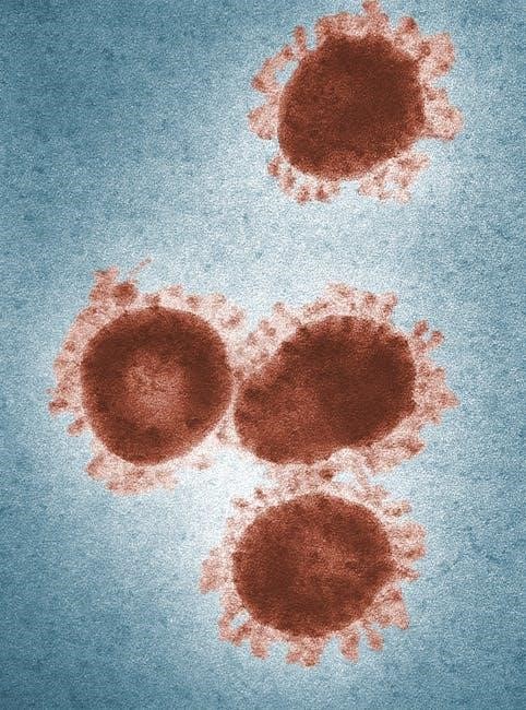
Microscopes are central to microbiology, as detailed in laboratory PDF manuals, enabling visualization of microorganisms. These instruments, crucial for both theory and application, allow observation of bacterial morphology, arrangement, and staining characteristics.
Different types, like brightfield, phase contrast, and potentially electron microscopes, offer varying levels of magnification and resolution. Understanding microscope operation, proper slide preparation, and focusing techniques are essential lab skills. Manuals emphasize their role in identifying unknown species and confirming the results of biochemical tests. Accurate microscopic observation forms the foundation of many microbiological analyses.
Sterilization and Disinfection Equipment
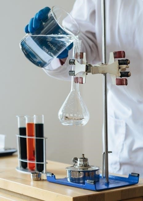
Sterilization and disinfection are paramount in microbiology, as outlined in laboratory PDF manuals, ensuring accurate results and lab safety. Autoclaves, utilizing high-pressure steam, are primary sterilization tools, eliminating all microbial life. Dry heat ovens offer an alternative method.
Disinfection, using chemical agents, reduces microbial load on surfaces. Manuals detail proper equipment operation, safety protocols, and validation of sterilization cycles. Maintaining a sterile environment prevents contamination and ensures reliable experimental outcomes. Universal precautions, including handwashing and lab coat use, are consistently emphasized for safe handling of potentially pathogenic cultures.
Incubators and Growth Chambers
Incubators and growth chambers, detailed in microbiology laboratory PDF manuals, provide controlled environments for microbial cultivation. These devices maintain optimal temperature, humidity, and often, CO2 levels, crucial for bacterial growth. Different species require specific conditions, necessitating precise control.
Manuals emphasize monitoring and calibration of these instruments to ensure consistent results. Growth observation is a key component, with documentation of colony morphology and growth rates. Proper incubation is vital for successful culturing and accurate biochemical testing, underpinning bacterial identification projects as described in various lab resources.
Aseptic Techniques & Safety Protocols
Microbiology lab manuals (PDF format) stress aseptic technique and universal precautions, including handwashing, lab coats, and proper handling of potentially pathogenic cultures.
Handwashing and Personal Protective Equipment
Microbiology laboratory manuals, frequently accessed as PDF documents, consistently emphasize the critical importance of rigorous handwashing with soap and water both before entering and upon leaving the laboratory environment. This foundational practice minimizes contamination risks.
Furthermore, these manuals mandate appropriate personal protective equipment (PPE). Specifically, they require fully covering footwear – prohibiting sandals or open-toe/heel shoes like clogs – and the consistent wearing of a lab coat while actively engaged in microbiological work.
These protocols, detailed in resources like the Pakpour & Horgan manual, are paramount for maintaining a safe and sterile workspace, protecting both the researcher and the integrity of experiments.
Sterilization Methods: Autoclaving, Dry Heat
Microbiology laboratory manuals, often available in PDF format, dedicate significant sections to sterilization techniques. These resources detail the principles and applications of both autoclaving and dry heat sterilization, essential for eliminating all microbial life.
Autoclaving, utilizing high-pressure steam, is frequently presented as a primary method for sterilizing media, glassware, and instruments. Conversely, dry heat sterilization, employing high temperatures for extended periods, is often recommended for materials unsuitable for autoclaving.
Understanding the nuances of each method, as outlined in comprehensive lab guides, is crucial for ensuring the sterility of laboratory materials and preventing contamination, upholding experimental validity.
Working with Microbial Cultures Safely
Microbiology laboratory theory, detailed in PDF manuals, emphasizes paramount safety protocols when handling microbial cultures. These guides consistently stress the importance of aseptic techniques and universal precautions to minimize exposure risks.

Essential practices include mandatory handwashing before and after lab work, appropriate personal protective equipment (PPE) like lab coats, and prohibiting open-toe footwear. Manuals from institutions like California State University explicitly outline these requirements.
Proper disposal of cultures and contaminated materials, alongside diligent disinfection of work surfaces, are also highlighted, ensuring a safe learning and research environment for all personnel.
Microbial Staining Techniques
PDF lab manuals detail staining procedures – simple, differential (like Gram stain), and special (acid-fast) – vital for visualizing and identifying microbial characteristics.
Simple Staining Procedures
Simple staining, as outlined in microbiology laboratory theory & application PDF manuals, employs a single dye to highlight microbial morphology. This foundational technique allows for basic observation of cell shape, size, and arrangement.
Common dyes include methylene blue, crystal violet, and safranin. These procedures are crucial introductory steps, providing a quick and easy method to assess sample purity and observe cellular structures before proceeding to more complex staining methods.
Lab manuals emphasize proper smear preparation and staining times for optimal results. While not differentiating between bacterial types, simple staining is a cornerstone of microscopic examination in microbiology.
Differential Staining: Gram Stain
Gram staining, a cornerstone technique detailed in microbiology laboratory theory & application PDF resources, differentiates bacteria based on cell wall structure. This differential staining method categorizes bacteria as Gram-positive (purple) or Gram-negative (pink/red).
The procedure involves crystal violet, Gram’s iodine, decolorization (typically with alcohol), and safranin counterstain. Manuals emphasize precise decolorization, crucial for accurate results. Gram-positive bacteria retain the crystal violet due to a thick peptidoglycan layer, while Gram-negative bacteria lose it during decolorization.
Understanding Gram stain results is vital for initial bacterial identification and guides further diagnostic testing, as highlighted in various lab manuals.
Special Staining: Acid-Fast Stain
Acid-fast staining, comprehensively covered in microbiology laboratory theory & application PDF guides, identifies bacteria with waxy mycolic acid in their cell walls, notably Mycobacterium species. These bacteria resist decolorization with acid-alcohol after being stained with carbolfuchsin.
The procedure often utilizes heat to aid carbolfuchsin penetration. Non-acid-fast bacteria are decolorized, appearing colorless, while acid-fast bacteria retain the red stain. This technique is crucial for diagnosing tuberculosis and other mycobacterial infections.
Lab manuals emphasize proper heat control and decolorization timing for accurate differentiation. Mastering acid-fast staining is essential for clinical microbiology and research.
Culturing and Media Preparation
Microbiology laboratory theory & application PDF resources detail diverse media types – broth, agar, selective, and differential – alongside inoculation methods like streak and spread plates.
Types of Culture Media (Broth, Agar, Selective, Differential)
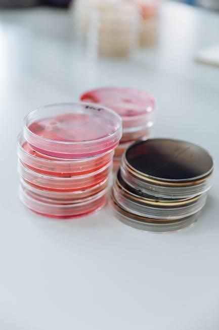
Microbiology laboratory theory & application PDF manuals extensively cover various culture media crucial for microbial growth. Broth media, liquid nutrient solutions, support large-scale cultivation. Agar media, solidified by agar, allows for visible colony formation and isolation.
Selective media contain components inhibiting certain microbial growth, isolating specific organisms. Conversely, differential media incorporate indicators revealing differing metabolic activities among microbes. These media formulations, detailed in lab manuals, are essential for identifying and characterizing bacterial species through observable growth patterns and biochemical reactions. Understanding these distinctions is fundamental to successful microbial culturing and analysis.
Methods of Inoculation (Streak Plate, Spread Plate)
Microbiology laboratory theory & application PDF resources detail essential inoculation techniques for isolating and cultivating microorganisms. The streak plate method, a cornerstone of microbiology, dilutes a sample across an agar surface, yielding isolated colonies. This technique, described in numerous lab manuals, allows for pure culture isolation.
Alternatively, the spread plate method evenly distributes a diluted sample across the agar surface, useful for quantifying bacterial populations. Both methods, fundamental to bacterial identification projects, require aseptic technique to prevent contamination. Mastering these inoculation techniques, as outlined in comprehensive manuals, is vital for accurate microbiological analysis.
Incubation Conditions and Growth Observation
Microbiology laboratory theory & application PDF guides emphasize the importance of controlled incubation for optimal microbial growth. Temperature, a critical factor, typically ranges from room temperature to 37°C, depending on the organism. Atmospheric conditions, like oxygen availability, also influence growth, necessitating aerobic or anaerobic incubators.
Careful observation of growth characteristics – colony morphology, color, and texture – is crucial for identification. Lab manuals detail these observations, linking them to specific bacterial species. Consistent monitoring and documentation, as highlighted in resources, are essential for accurate results and successful bacterial identification projects.

Biochemical Testing for Identification
Microbiology laboratory theory & application PDF resources detail tests like catalase, oxidase, and sugar fermentation. These identify bacteria based on metabolic capabilities, aiding species determination.
Catalase and Oxidase Tests
Catalase tests, detailed in microbiology laboratory theory & application PDF manuals, determine the presence of catalase enzyme, breaking down hydrogen peroxide. A positive result shows bubble formation, indicating the organism possesses catalase. Conversely, oxidase tests identify cytochrome c oxidase, crucial for electron transport.
These tests, fundamental in bacterial identification, utilize reagents to detect enzymatic activity. Positive oxidase reactions display a color change, indicating enzyme presence. Manuals from institutions like California State University emphasize these tests’ importance in differentiating bacterial species based on metabolic pathways. Accurate interpretation, guided by lab manuals, is vital for correct identification.
Sugar Fermentation Tests
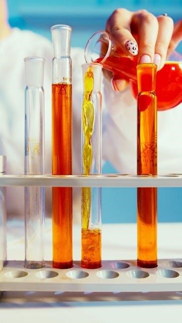
Sugar fermentation tests, comprehensively outlined in microbiology laboratory theory & application PDF resources, assess a bacterium’s ability to ferment specific sugars. These tests utilize phenol red indicator, revealing acid production (yellow color) or alkalinity (red color). Manuals detail inoculating bacteria into sugar-containing broths, observing for gas production via Durham tubes.
These tests are crucial for differentiating bacteria based on metabolic capabilities, as highlighted in various lab manuals. Positive fermentation indicates acid production, while negative results show no change. Understanding these principles, detailed in resources like Pearson’s Microbiology A Laboratory Manual, is vital for accurate bacterial identification.
IMViC Tests (Indole, Methyl Red, Voges-Proskauer, Citrate)
IMViC tests – Indole, Methyl Red, Voges-Proskauer, and Citrate – are a series of biochemical tests detailed in microbiology laboratory theory & application PDF manuals. These tests differentiate Gram-negative bacteria based on differing metabolic pathways. Each test utilizes specific reagents to detect unique enzymatic activities;
Lab manuals, like those from introductory microbiology courses, explain procedures for each test. For example, the Indole test detects tryptophan breakdown. Mastering these tests, as emphasized in resources like Pearson’s lab manuals, is essential for accurate bacterial identification and characterization within a microbiology laboratory setting.
Bacterial Identification Projects & Reporting
Microbiology laboratory theory & application PDF resources emphasize analyzing test results to identify unknown bacteria. Reports detail procedures, observations, and conclusions, crucial for project assessment.
Analyzing Test Results
Analyzing results from biochemical tests, staining procedures, and growth observations is central to a microbiology laboratory theory & application approach. PDF manuals guide students through interpreting data from catalase, oxidase, sugar fermentation, and IMViC tests.
Careful examination of these outcomes, alongside morphological characteristics revealed by staining, allows for the construction of a differential table. This table systematically compares observed results with known characteristics of various bacterial species.
The process requires critical thinking to discern patterns and eliminate possibilities, ultimately leading to a tentative identification. Accurate record-keeping, as emphasized in lab reports, is paramount for reliable analysis.
Identifying Unknown Bacterial Species
Identifying unknown bacterial species relies heavily on the principles detailed in microbiology laboratory theory & application resources, often available as PDF manuals. This process integrates data from all performed tests – staining, biochemical assays, and growth characteristics.
Students systematically compare their accumulated results against established identification schemes, like dichotomous keys or comprehensive tables found within lab manuals.
The goal is to narrow down possibilities until a single, most probable species is determined. Confirmation may involve further testing or referencing specialized databases. A well-structured laboratory report, documenting the entire process, is crucial for demonstrating accurate identification.
Laboratory Report Structure and Content
Microbiology laboratory theory & application PDF manuals emphasize a standardized report structure. Typically, reports begin with an introduction outlining the experiment’s purpose and relevant background theory; A detailed materials and methods section follows, documenting procedures precisely.
Results are presented clearly, often using tables and figures, avoiding interpretation at this stage. The discussion section interprets findings, relating them to the initial hypothesis and existing literature.